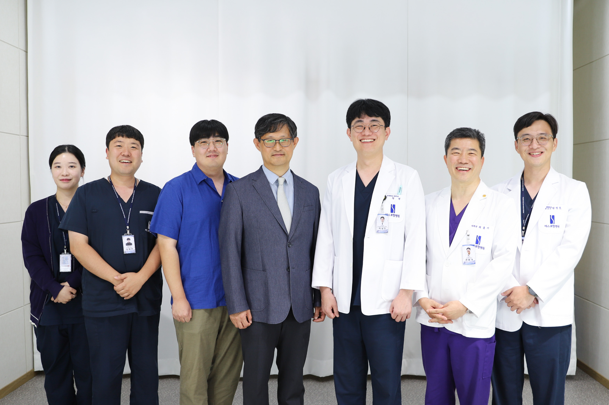
한동대학교 AI컴퓨터전자공학부 딥러닝 연구실(지도교수: 김인중)은 에스포항병원과 협력해 척추 수술 후 X-ray 영상 분석을 위한 생성형 AI 기술을 개발했다. 이번 연구는 영상의학 인공지능 분야의 국제학술지 『Radiology: Artificial Intelligence』에 게재가 확정됐다. 해당 학술지는 북미영상의학회(RSNA)가 발행하며, 2025년 기준 피인용지수(Impact Factor)가 20.1로 영상의학·핵의학·의료영상 분야 217개 학술지 중 1위에 해당한다.
이번 연구에는 최진영 박사과정 대학원생과 AI컴퓨터전자공학부 학부과정 김석원·송승언·박인수 학생이 참여해 AI 모델 개발과 실험을 수행했으며, 에스포항병원 의료진은 임상 데이터 구축과 의학적 검증에 참여했다. 이번 연구는 대학과 지역 의료기관이 협력해 실제 임상 현장의 문제를 해결하기 위한 의료 AI 기술을 개발했다는 점에서 의미가 있다.
척추 유합술 후 X-ray 영상에는 금속 나사와 케이지 등의 임플란트가 척추체를 가려 기존 AI 기술로 척추의 형태와 정렬 상태를 분석하기 어려운 문제가 있었다. 연구팀은 임플란트가 없는 정상 X-ray 영상으로 생성형 AI를 학습시킨 뒤, 원본 영상과 AI 복원 영상의 차이를 활용해 임플란트 영역을 찾아 제거하고 주변 뼈의 형태를 복원하는 ARNAI(Artifact Removal Network based on Autoencoding and Inpainting) 기술을 개발했다. 이를 통해 복원된 영상에서 척추 분할과 정렬 지표를 자동으로 분석하는 RSM(Restore, Segment, and Measure) 프레임워크를 구축했다.
연구 결과, 자동 분석이 불가능했던 기각 영상이 외부 검증에서 65.45% 감소했으며, 요추 4-5번 분절의 각도 측정 오차도 내부 검증에서 약 70% 감소했다. 김인중 교수는 “임플란트가 포함된 학습 데이터를 따로 모으지 않고도 수술 후 영상 분석이 가능하다는 점을 보인 것이 이번 연구의 핵심”이라며 “한동대가 지역의 병원과 협력을 통해 의료 AI를 개발하고 의료영상 분야의 주요 국제학술지로부터 연구성과를 인정받았다는 점에서 의미가 있다”고 말했다.
관련 기사: "한동대·에스포항병원, 척추 엑스레이 분석 인공지능 개발", 한국일보
https://www.hankookilbo.com/news/article/A2026092809520002114
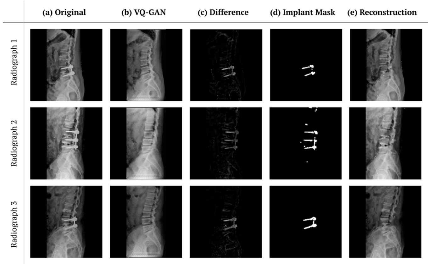
< ARNAI가 수술 후 X-ray 영상으로부터 임플란트를 거하는 과정 >
The Deep Learning Lab at the School of Artificial Intelligence, Computer and Electrical Engineering (AICE) at Handong Global University, led by Professor Injung Kim, has developed a generative AI technology for analyzing postoperative spine X-ray images in collaboration with Pohang Stroke and Spine Hospital. The study has been accepted for publication in Radiology: Artificial Intelligence, an international journal in the field of medical imaging published by the Radiological Society of North America (RSNA). With an Impact Factor of 20.1 in 2025, the journal ranked first among 217 journals in the fields of Radiology, Nuclear Medicine, and Medical Imaging.
The study involved Ph.D. student Jinyoung Choi and undergraduate students Seokwon Kim, Seungeon Song, and Insu Park from AICE, who contributed to AI model development and experiments. Medical staff at Pohang Stroke and Spine Hospital contributed to clinical data construction and medical validation. The study demonstrates how collaboration between a university and a regional medical institution can address real-world clinical challenges through medical AI research.
Postoperative X-ray images often contain metal implants such as screws and cages, making it difficult for AI systems to accurately analyze spinal structure and alignment. The research team developed ARNAI (Artifact Removal Network based on Autoencoding and Inpainting), which identifies and removes implant regions by comparing original X-ray images with AI-reconstructed images trained on normal X-rays without implants. The team also developed the RSM (Restore, Segment, and Measure) framework to perform spinal segmentation and alignment measurement using the reconstructed images.
The results showed a 65.45% reduction in images rejected as unsuitable for automated analysis in external validation, while the mean absolute error in measuring the L4–L5 segment angle decreased by approximately 70% in internal validation. Professor Injung Kim stated, “The key point of this study is that it demonstrated the feasibility of analyzing postoperative images without separately collecting training data containing implants.” He added that the research is meaningful in demonstrating how Handong Global University and a regional hospital collaborated to develop medical AI and gained recognition from a leading international journal in medical imaging.
Related Article: "한동대·에스포항병원, 척추 엑스레이 분석 인공지능 개발", 한국일보
https://www.hankookilbo.com/news/article/A2026092809520002114


한동대학교 AI컴퓨터전자공학부 딥러닝 연구실(지도교수: 김인중)은 에스포항병원과 협력해 척추 수술 후 X-ray 영상 분석을 위한 생성형 AI 기술을 개발했다. 이번 연구는 영상의학 인공지능 분야의 국제학술지 『Radiology: Artificial Intelligence』에 게재가 확정됐다. 해당 학술지는 북미영상의학회(RSNA)가 발행하며, 2025년 기준 피인용지수(Impact Factor)가 20.1로 영상의학·핵의학·의료영상 분야 217개 학술지 중 1위에 해당한다.
이번 연구에는 최진영 박사과정 대학원생과 AI컴퓨터전자공학부 학부과정 김석원·송승언·박인수 학생이 참여해 AI 모델 개발과 실험을 수행했으며, 에스포항병원 의료진은 임상 데이터 구축과 의학적 검증에 참여했다. 이번 연구는 대학과 지역 의료기관이 협력해 실제 임상 현장의 문제를 해결하기 위한 의료 AI 기술을 개발했다는 점에서 의미가 있다.
척추 유합술 후 X-ray 영상에는 금속 나사와 케이지 등의 임플란트가 척추체를 가려 기존 AI 기술로 척추의 형태와 정렬 상태를 분석하기 어려운 문제가 있었다. 연구팀은 임플란트가 없는 정상 X-ray 영상으로 생성형 AI를 학습시킨 뒤, 원본 영상과 AI 복원 영상의 차이를 활용해 임플란트 영역을 찾아 제거하고 주변 뼈의 형태를 복원하는 ARNAI(Artifact Removal Network based on Autoencoding and Inpainting) 기술을 개발했다. 이를 통해 복원된 영상에서 척추 분할과 정렬 지표를 자동으로 분석하는 RSM(Restore, Segment, and Measure) 프레임워크를 구축했다.
연구 결과, 자동 분석이 불가능했던 기각 영상이 외부 검증에서 65.45% 감소했으며, 요추 4-5번 분절의 각도 측정 오차도 내부 검증에서 약 70% 감소했다. 김인중 교수는 “임플란트가 포함된 학습 데이터를 따로 모으지 않고도 수술 후 영상 분석이 가능하다는 점을 보인 것이 이번 연구의 핵심”이라며 “한동대가 지역의 병원과 협력을 통해 의료 AI를 개발하고 의료영상 분야의 주요 국제학술지로부터 연구성과를 인정받았다는 점에서 의미가 있다”고 말했다.
관련 기사: "한동대·에스포항병원, 척추 엑스레이 분석 인공지능 개발", 한국일보
https://www.hankookilbo.com/news/article/A2026092809520002114
< ARNAI가 수술 후 X-ray 영상으로부터 임플란트를 거하는 과정 >
The Deep Learning Lab at the School of Artificial Intelligence, Computer and Electrical Engineering (AICE) at Handong Global University, led by Professor Injung Kim, has developed a generative AI technology for analyzing postoperative spine X-ray images in collaboration with Pohang Stroke and Spine Hospital. The study has been accepted for publication in Radiology: Artificial Intelligence, an international journal in the field of medical imaging published by the Radiological Society of North America (RSNA). With an Impact Factor of 20.1 in 2025, the journal ranked first among 217 journals in the fields of Radiology, Nuclear Medicine, and Medical Imaging.
The study involved Ph.D. student Jinyoung Choi and undergraduate students Seokwon Kim, Seungeon Song, and Insu Park from AICE, who contributed to AI model development and experiments. Medical staff at Pohang Stroke and Spine Hospital contributed to clinical data construction and medical validation. The study demonstrates how collaboration between a university and a regional medical institution can address real-world clinical challenges through medical AI research.
Postoperative X-ray images often contain metal implants such as screws and cages, making it difficult for AI systems to accurately analyze spinal structure and alignment. The research team developed ARNAI (Artifact Removal Network based on Autoencoding and Inpainting), which identifies and removes implant regions by comparing original X-ray images with AI-reconstructed images trained on normal X-rays without implants. The team also developed the RSM (Restore, Segment, and Measure) framework to perform spinal segmentation and alignment measurement using the reconstructed images.
The results showed a 65.45% reduction in images rejected as unsuitable for automated analysis in external validation, while the mean absolute error in measuring the L4–L5 segment angle decreased by approximately 70% in internal validation. Professor Injung Kim stated, “The key point of this study is that it demonstrated the feasibility of analyzing postoperative images without separately collecting training data containing implants.” He added that the research is meaningful in demonstrating how Handong Global University and a regional hospital collaborated to develop medical AI and gained recognition from a leading international journal in medical imaging.
Related Article: "한동대·에스포항병원, 척추 엑스레이 분석 인공지능 개발", 한국일보
https://www.hankookilbo.com/news/article/A2026092809520002114